
Embryology Atlas: From Fertilization to Blastocyst
Welcome to the VCRM Embryology Atlas. Take a visual journey through the microscopic stages of human embryonic development—from mature oocyte retrieval and ICSI fertilization to day-by-day cell division and blastocyst hatching in our state-of-the-art cleanroom IVF laboratory.
IVF Lab Excellence
Our Class 100 cleanroom embryology lab is directed by board-certified embryologists and Dr. Fady Sharara.
Schedule Consultation (571) 443-8322Blastocyst Development: The First 6 Days of Life
In human reproduction, fertilization triggers an intricate series of cellular divisions. In the VCRM embryology laboratory, our team monitors each embryo's morphokinetic milestones—assessing timing, cleavage symmetry, cell number, and blastocoel expansion.
Watch this educational time-lapse video demonstrating the continuous development of a fertilized human zygote into an expanded Day 5 blastocyst:
Normal Embryo Development: Stage by Stage
Below are high-magnification inverted microscope photographs showing the ideal progression of healthy human gametes and embryos during an IVF / ICSI cycle at VCRM:
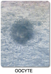
1. Mature Oocyte (MII)
A healthy mature Metaphase II human egg. Note the clear, spherical cytoplasm, smooth zona pellucida, and distinct first polar body indicating readiness for fertilization.
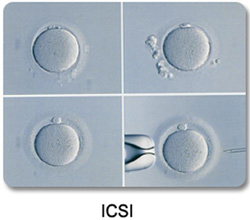
2. ICSI Procedure
A single immobilized sperm is delicately injected into the mature oocyte using an ultra-fine glass micropipette while held firmly by gentle suction.
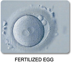
3. Normal Fertilization (2PN)
16–18 hours post-insemination, two distinct pronuclei (2PN) are visible—one carrying maternal DNA and one carrying paternal DNA—along with two polar bodies.
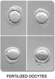
4. Synchronous Zygote Cohort
Multiple fertilized 2PN zygotes developing synchronously in individualized microwells within our advanced low-oxygen incubators.
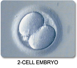
5. 2-Cell Cleavage Embryo
The first mitotic cellular division produces two evenly sized blastomeres with clear cytoplasm and minimal fragmentation.
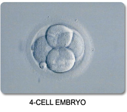
6. 4-Cell Stage Embryo
Approximately 44–48 hours post-retrieval, the embryo divides into 4 symmetrical blastomeres within an intact protective zona pellucida.
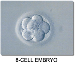
7. 8-Cell Cleavage Embryo
Day 3 benchmark showing 8 cohesive cells. Shortly after this stage, cellular compaction begins as the embryo transitions into a morula.
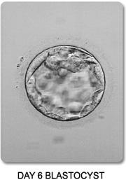
8. Fully Expanded Blastocyst
Contains a fluid-filled blastocoel cavity, a tight inner cell mass (which forms the fetus), and an outer trophectoderm layer (which forms the placenta).
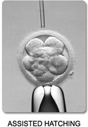
9. Assisted Hatching
A microscopic opening is created in the outer shell (zona pellucida) using a precision infrared laser, facilitating embryo hatching and implantation or genetic biopsy.
Abnormal Embryo Morphology & Variations
Not all oocytes or embryos develop normally. Morphological variations, fragmentation, abnormal fertilization, and cell arrest provide vital diagnostic insight into gamete quality, chromosomal integrity, and cycle optimization:
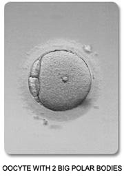
1. Dysmorphic Oocyte
Oocyte displaying irregular outer morphology and expanded perivitelline space, which may reflect diminished cytoplasmic maturity.
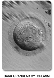
2. Dark Granular Cytoplasm
Central cytoplasmic granularity and darkness, often associated with elevated oxidative stress or mitochondrial alterations.
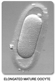
3. Elongated / Oval Oocyte
Non-spherical, elongated zona pellucida and ooplasm that may present technical challenges during micro-manipulation or ICSI.
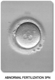
4. Abnormal 3PN Fertilization
Three pronuclei (3PN) visible instead of two. This triploid zygote is chromosomally abnormal and cannot be transferred.
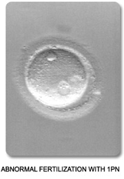
5. Multi-Pronuclear Zygote
Aberrant pronuclear formation indicating failed maternal second meiotic division or polyspermic fertilization.
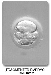
6. Day 2 Fragmented Embryo
Small non-nucleated membrane-bound cytoplasmic fragments surrounding uneven blastomeres during early cleavage.
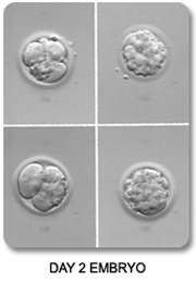
7. Heavy Cleavage Fragmentation
> 30% cytoplasmic volume lost to cellular fragmentation, which can reduce blastocyst conversion rates.
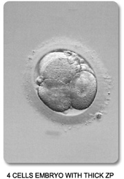
8. 4-Cell with Thickened Zona
Asymmetrical cleavage accompanied by an abnormally thick outer zona pellucida that often benefits from laser-assisted hatching.
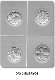
9. Fragmented Day 3 Embryo
Irregular cellular division at Day 3 with prominent debris interfering with cell-to-cell junctions necessary for compaction.
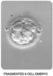
10. Fragmented 8-Cell Stage
Eight blastomeres obscured by extensive fragmentation. Many such embryos self-correct or stall before blastulation.
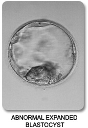
11. Degenerated / Arrested Blastocyst
A collapsed blastocoel cavity with sparse, apoptotic cells and absent inner cell mass, indicating developmental arrest.
VCRM Embryology Laboratory Standards
Embryo quality is deeply influenced by the laboratory environment. Under the direction of Chad A. Johnson, PhD, HCLD and Dr. Fady Sharara, the VCRM IVF Laboratory adheres to the most stringent international standards:
Class 100 Air Quality
Positive pressure cleanroom with active VOC carbon filtration and HEPA filters eliminating airborne toxins.
Tri-Gas Benchtop Incubators
Individualized low-oxygen chambers that mimic physiological fallopian and uterine conditions for optimal growth.
Ultra-Rapid Vitrification
Flash freezing that yields > 98% blastocyst survival rates for Frozen Embryo Transfer (FET).
Frequently Asked Questions About Embryo Development
Can an embryo with fragmentation still result in a healthy baby?
Yes. Mild to moderate fragmentation (Grade 1 or 2, < 15–20%) is very common and embryos frequently self-correct as they develop to the blastocyst stage. Many healthy babies have been born from embryos that exhibited mild early cleavage fragmentation.
Why do some embryos stop growing (arrest) between Day 3 and Day 5?
During the first three days, the embryo relies primarily on maternal RNA stored within the egg. On Day 3, the embryo's own genome activates (embryonic genome activation). If there are significant paternal or maternal chromosomal abnormalities or mitochondrial deficits, development may arrest before blastocyst formation.
What is the difference between Day 3 cleavage and Day 5 blastocyst transfers?
Day 5 blastocysts have demonstrated their ability to survive genome activation and compaction. Transferring a blastocyst provides higher implantation rates and allows precise cell biopsy for PGT-A/PGT-M genetic testing.
How does laser assisted hatching help embryos implant?
Before an embryo can implant in the uterine lining, it must "hatch" out of its outer shell (the zona pellucida). Assisted hatching thins or breaches this shell, which is especially beneficial for frozen embryo transfers, older maternal age, or embryos with thickened shells.

Dr. Fady I. Sharara, M.D., FACOG & Chad A. Johnson, PhD
Medical Director & Laboratory Leadership at Virginia Center for Reproductive Medicine, specializing in advanced embryology, micro-manipulation, and high-complexity IVF care.
Experience Boutique Embryology Care at VCRM
Schedule your personal consultation with Dr. Fady Sharara to learn more about our IVF protocols and world-class laboratory technology.